Product information “Foot – Deep plantar structures”
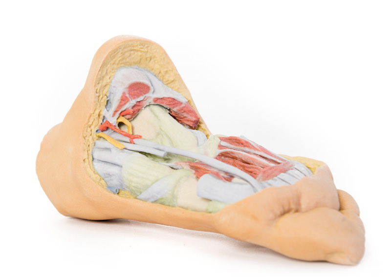
This 3D printed specimen provides a view of deep plantar structures of a right foot. Medially, the cut edge of the great saphenous vein is visible within the superficial fascia, just anterior to the cut edges of the medial and lateral plantar arteries and nerves overlying the insertion of the tibialis posterior muscle. The superficial fascias, the plantar aponeurosis, and superficial musculature have been removed to expose the deep (third layer) of musculature.
Search
Enquiry Line: 01 803 8688
Welcome To Medstore Medical - Thousands Of Products - Nationwide - Worldwide

