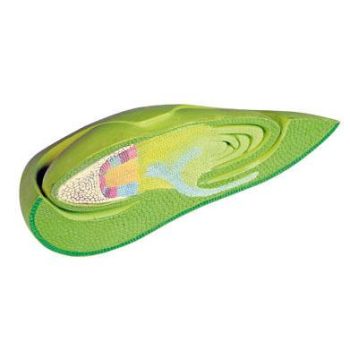
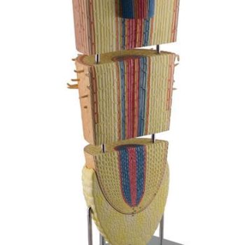
Description
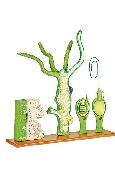
This 4-part model shows longitudinal and cross sectional views of the hydra, highlighting significant anatomical features, including the ectoderm, mesoglea, coelenteron, male and female egg cells, buds and mouth opening. The cross sectional block model, about 250X life size, shows various cellular structures, including nematoblasts, epithelial cells, sensory cells, interstitial cells and the nerve network. An additional block model shows the anatomy of a highly enlarged cnidoblast cell.
Search
Enquiry Line: 01 803 8688
Welcome To Medstore Medical - Thousands Of Products - Nationwide - Worldwide

